Our Variability Restoration Framework
Welcome to Part 2 of this five-part series. In Part 1, we covered the overarching neuromechanical features of our movement system that offer us a general insight into how our anatomy is designed to help us manage & distribute forces.
Here, we will dive deeper into the general framework that we use to guide the restoration of variability within our movement system. This highlights how we use muscle & joint behavior to guide our thought process as we work through assessment & rehabilitation.
Movement variability
Variability is an intrinsic characteristic of our movement system that was once viewed as an unwanted feature that should be minimized. Not anymore! From a movement perspective, variability represents the movement of our body segments that occur between each repetition of a task, and this gives our system many options to share & distribute forces that act on our system.
Movement variability is often classified according to two components:
- Variability in the outcome / goal of a task
- Variability of the elements (muscles & joints) that represent our anatomy
For example, if you were to point to a small target on a wall in front of you, there are many combinations of upper limb muscle & joint movement patterns that will enable you to accurately point to that target. This would represent increased variability of the elements, but low variability of the goal / outcome (small target).
In the health & fitness space, these two components are often confused, especially in the search for “optimal” movement. The idea is that there is an optimal way that our muscles & joints will organize themselves to achieve the task outcome. However, in the example above, we can appreciate that there are multiple ways (muscle & joint patterns) to achieve an optimal outcome. It’s much easier to assess if an optimal outcome has been achieved, but because our movement system is so complex, it’s impossible to determine an optimal behavior of the elements of our anatomy.
Variability within our movement system changes in the presence of pain. Generally, individuals move with less range (less muscular & joint variability), move at lower speeds, and move with more muscle activity. When movement is related to pain, and it’s influenced within the treatment/rehabilitation process, we generally see that range & speed of movement increase, as well being more relaxed (Wernli, 2020)
Within our approach at IKN, we categorize rehabilitation into three categories:
- Restoring muscular & joint variability
- Symptom & activity modification
- Gradual exposure to meaningful activities
We will discuss these three categories more in Part 4 of this series, but here we will explore the framework that we use to restore variability within the movement system.
Our Variability Framework
Variability of our muscles & joints is often referred to as “coordinative variability.” Research surrounding coordinative variability aims to characterize the interactions across muscles & joints, and understand how these interactions influence our systems ability to manage & share forces. How do they change in the presence of injury and/or pain? How can we use this information to guide our treatment and rehab?
To simplify this concept, we like to use muscle & joint behavior as our ultimate guide to assess variability. Remember, this is not the only feature we look at, but it’s an important one to guide where, when, and how we load / apply stress during rehab.
Our restoring variability framework involves:
- Identifying current muscular & joint/segmental variability
- Organizing our loading/progressions/regression around restoring variability / increasing movement options
We DO NOT organize our rehab relative to “ideal” or “optimal” movement, but towards a representation of greater variability. There is a huge difference between these two thought processes! One believes in “faulty” or “dysfunctional” movement, while the other sees them as normal variations in movement (variability of the elements).
Additionally, we couple this muscular & joint/segmental framework with other crucial biopsychosocial features to guide rehabilitation.
Below, we will expand on the local and global features of muscular and joint variability.
Muscular features
To simplify muscular behavior, we can view it from two separate perspectives:
- Local muscular variability: this represents the local capacity of muscles to manage & produce forces with concentric, isometric, and eccentric behaviors. This can also be characterized as INTRA-muscular variability / within-muscle variability.
- Global muscular variability: this represents the capacity of muscles to distribute forces between neighboring muscular tissues & help share forces over broader surface areas. This can also be characterized as INTER-muscular variability / between-muscle variability.
Joint / Segmental features
Joint complexes across our anatomy are simply representations of interacting segments. Within our approach, we use a segmental-based analysis vs a joint-based analysis.
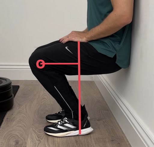
For example, in both images shown, we can clearly see that the knee is flexed. However, characterizing this as “knee flexion
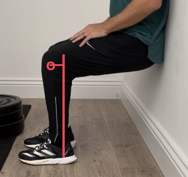 ” does not paint a clear picture of how forces will be acting on the muscular tissues surrounding the “knee joint.” From a segmental perspective, we need to appreciate the interaction / angles between the femoral and tibial segments (and of coursethe patella).
In image the bottom image, we can see that the femur is still parallel to the ground, but the tibia is more flexed (a forward shin angle). This means that there will be greater load through the distal quadriceps because gravity will try to create more tibial flexion, which will require more force production demand through the distal quadriceps. In the top image, the tibia is more vertical, which reduces the demand on the distal quadriceps.
You could ask, “well what if we just say that the knee is more flexed in the bottom image?” This still doesn’t help, because it doesn’t tell us how that flexion is occurring. Is it occurring through more tibial flexion or more femoral flexion? We believe that we need to shift towards using this segmental-based analysis to paint a clearer picture of how forces are acting on the muscular tissues.
From a local segmental perspective, there are many ways that segments can express variability. Within coordinative research, the capacity of neighboring segments to dissociate from each other is one of the key representations studied.
This represents the ability of neighboring segments to move independently, or in opposite directions. In coordinative research, they refer to the capacity of segments to move in opposite directions as “antiphase” patterns, and when they move in the same direction, they refer to them as “inphase” patterns.
Case insights
We will keep it simple here, and just share two clinical representations of low muscular & joint variability.
Neck variability = head segment & thoracic spine / rib cage interaction
You ask your patient to flex their head, and as they begin to flex their head, you see them quickly flex their thoracic spine / rib cage to assist “neck” flexion. This may be secondary to an inability to facilitate eccentric behavior through the upper cervical extensors, or perhaps they are sensitive to neck flexion, and need to share flexion into the thoracic spine. This represents an “inphase” strategy, where the head & thoracic spine move in the same direction (flexing together), i.e., the thoracic spine can’t stay relatively extended as they concentrate flexion through the head / neck. This is not a “bad” thing, but just the strategy they are currently using.
Elbow variability = humeral segment & forearm (radius & ulna) interaction
You have a patient with lateral elbow pain, and you are assessing their active forearm pronation ability. But, you notice that as they pronate their forearm, their humerus quickly internally rotates to assist this forearm pronation. This represents that they likely don’t have the capacity to pronate the forearm relative to the humerus, as both are internally rotating to get the palm of their hand to face down (inphase pattern). This may be secondary to an inability to express eccentric behavior through the supinator muscle, or because they are expressing increased muscle guarding around the elbow, leading to a lack of dissociation.
These representations are not good or bad, but they help to give us an insight into the current level of variability being expressed between segments. Using these representations, we can consider what we may need to do from a rehabilitation standpoint.
Research
Seay et al, looked at these coordinative variability representations understand muscle and joint behavior in three groups of runners:
1 ) Runners with current low back pain
2 ) Runners who have recovered from low back pain
3 ) Runners who have never experienced low back pain
What they found was that the capacity to express dissociation / antiphase patterns between the trunk and pelvis in the transverse plane was:
– Greatest in those never injured (higher variability between segments)
– Smallest in those with current back pain (lower variability)
– In between for those that have recovered from previous back pain (hmm)
What we find most interesting about this is that in the group that had recovered from pain, there still existed a reduction in variability between those segments (trunk relative to pelvis), which may potentially reflect a continued challenge with sharing forces / loads across the trunk & pelvic region.
Perhaps, this persistent lack of variability in those who recovered represents that they are still expressing some of the movement adaptations they expressed when they were experiencing pain. Perhaps, greater focus was placed on organizing rehab around their symptoms vs integrating this with a focus on restoring variability back to the system?
For us, appreciating muscle & joint variability should be a crucial component of your assessment & rehabilitation framework, because many use pain as the only variable to guide recovery, when we really should be using multiple variables to guide things. Using pain to guide treatment / rehab is important in certain situations, but it should never be the only variable you use as guidance.
In conclusion..
Phew! This was a long blog, with a lot of information! For those of you that made it this far – thank you!
Variability is a complex topic, but we try to simplify it as much as we can when assessing the current movement capacities of our patients to help guide what we do from a rehabilitation standpoint.
Remember, using the variability of our movement system is just one component of what you should use to guide your thought process. Other important components include:
- Your patients’ beliefs & fears
- Their goals
- Symptom behavior (aggravating & easing factors)
- Prior injuries / pain experiences
- Prior treatment / rehab (what has worked / has not worked in the past?)
- Sensitivity levels (how sensitive are they to load / stress?)
- Diagnosis (mechanical vs non-mechanical?)
- And more….
Up next is part 3, where we will use the information covered in part 1 & 2 to guide our assessment process, i.e., how we identify the current variability of the system & integrate it with these other features to guide appropriate entry points to loading / rehab. Thanks again for reading!
References
Wernli K, Smith A, Coll F, Campbell A, Kent P, O’Sullivan P. From protection to non-protection: A mixed methods study investigating movement, posture and recovery from disabling low back pain (2022)
Seay JF, Van Emmerik RE, Hamill J. Low back pain status affects pelvis-trunk coordination and variability during walking and running (2011)