Foot Biomechanics Made Simple
When individuals experience foot and ankle limitations, the knee-jerk reaction is to address the ankle, which represents the interaction between the tibia/fibula and the talus. However, very few consider the variability WITHIN the foot itself (INTRA-foot variability), which involves appreciating how the rearfoot, midfoot, and forefoot interact with each other.
In our Level 1 & 2 courses, we categorize these interactions within the foot as follows, and we’ll also add some questions that guide our thought process when assessing them:
- Rearfoot: the interaction between the tibia/fibula, talus, calcaneus & tarsal bones
- Does the ankle have normal sagittal plane range of motion?
- Does the calcaneus have the capacity to invert & evert?

- Can the calcaneus express these motions during the appropriate sagittal plane motion? I.e., plantar flexion & dorsiflexion
- Midfoot: the interaction between the tarsal bones and the metatarsals
- Can the tarsal bones lift & lock to facilitate supination?
- Can the tarsal bones drop & unlock to facilitate pronation?
- Are the metatarsals facilitating this locking & unlocking of the tarsal bones?
- Forefoot: the interaction between the metatarsals and toes
- Can the toes (particularly the big toe) flex & extend?
- Can the metatarsals plantar flex & dorsiflex?
Let’s describe these interactions below within the context of facilitating greater supination and pronation capacities at the foot, and lets focus on the midfoot & forefoot interaction.
Supination & Pronation: A Forefoot View
To facilitate supination, the first metatarsal needs to express plantar flexion. This is shown in the image, where you can see a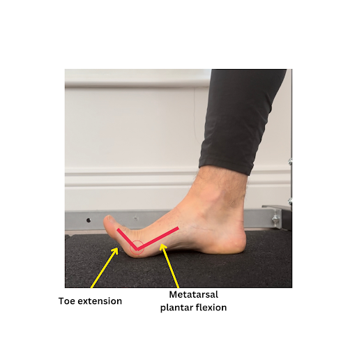 lifting of the metatarsal proximally, and a dropping of the metatarsal distally.
What’s also key to understand here is how this metatarsal behavior couples with toe motion. Metatarsal plantar flexion as you see here will couple with toe extension.
Try this: with your foot flat on the ground, extend your toes, and what you should see is a lifting of the medial arch. This lifting of the arch and the tarsal bones is generated largely through the plantar flexion of the metatarsal.
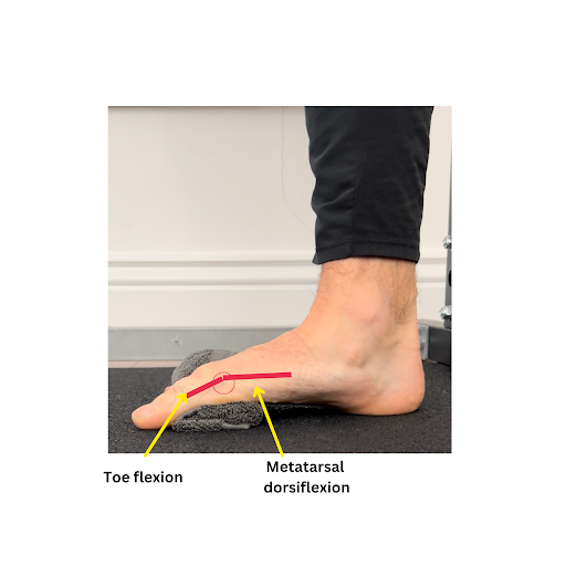
The opposite metatarsal & toe motion coupling is expressed to facilitate foot pronation. In the image shown, you can see a dorsiflexion of the metatarsal, where the metatarsal lifts distally and drops proximally. This dropping proximally enables the tarsal bones to drop, unlock, and facilitate a lengthening of the medial arch.
You can also appreciate from the image that this occurs in conjunction with toe flexion, which is key to enable the lifting of the metatarsal head distally.
So why is this important?
Like we mentioned at the beginning, variability within the foot is underappreciated when it comes to restoring the capacity to share forces across the foot and ankle. Typically, more focus is directed at the ankle/talocrural joint to restore variability to the distal end of the lower limb.
To keep it simple:
If you’re trying to facilitate foot supination? Consider the capacity of the metatarsals to plantar flex and the toes to extend.
If you’re trying to facilitate foot pronation? Consider the capacity of the metatarsals to dorsiflex and the toes to flex.